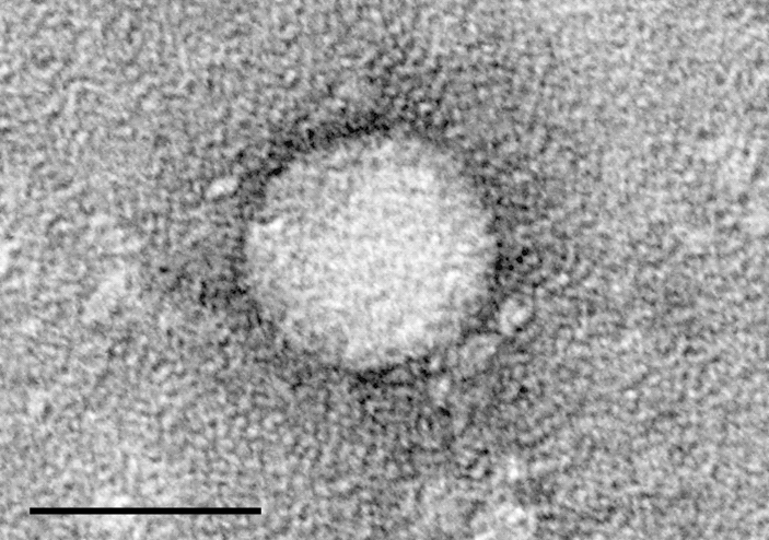
Hep C is disease caused by a virus; to understand the disease you have to understand the virus - a coxsackievirus - a positive-sense single-stranded RNA virus ("ssRNA(+)") in the family genus Hepacivirus, Genus Hepacivirus, species Hepacivirus C hence the name "Hepatitis C". "Hepatitis" means "infection of the liver" and is an archaic term. Hepaciviris is the name of the virus, "hepatitis" is the name of the disease, and there are three forms, A B and C all caused by different and fairly unrelated viruses. Hep C is abbreviated "HCV" for "Hep C Virus".
These three Hep viruses do not actually have that much in common, they are not in the same genus and or even in the same family, They just happen to be viruses of various types that happen to have one thing in common: they infect the liver. That is they are not thee forms of the same virus they are three different viruses that all happen to attack the liver and produce a fairy profound liver disease state.
Just as larger parasites are very host specific - the rat lung tapeworm, predictably, can only live in rat lung' dogs get canine distemper; cats and rats have their own species of fleas and so forth and so on - viruses too can be very host specific. Within the virus kingdom, Polio is notorious for residing in nerve ganglia, the cause of rare but notorious paralysis, Ebola "hides" in cartilage, polio reproduces in the gut, thus Hepatitis inducing vital pathogens A, B and C are simply three unrelated viruses that can each cause disease state in the liver as is the merely one of the parts of the body these viruses favor the others are pancreas and lymphatic system.
Hepatovirus A is a species of virus in the order Picornavirales in the family Picornaviridae and is the type species of the genus Hepatovirus. Humans and vertebrates serve as natural hosts.
Hepatitis B virus, abbreviated HBV, is a partially double-stranded DNA virus,[1] a species of the genus Orthohepadnavirus and a member of the Hepadnaviridae family of viruses.
Synonym: Hepatitis C virus famly Flaviviridae
Remediating a virally induced hepatitis requires understanding of the specific pathogen.
. All coxsackie viruses are selenium dependant.[1] You can not clear them without decent levels of selenium in the blood, two brazil nuts a day has been shown to raise it enough and it takes about 12 weeks.[2] to start working. It should measurable decline after that. Niacin and C would indeed help. Other Coxsackie virus disease also deplete the body of glutamine, cysteine and tryptophan. Tryptophan is made from 5-HTP, which is made from niacin, albeit inefficiently. Consider supplementing with this. Tryptophan is high in beef ad not much else. Small amounts daily would help. Cysteine is higher in cheese. Harald Foster said on BBC radio that "a cheeseburger made with brazil nut flour" would procice the missing raw material the immune system needs to clear the virus.
[1] "and more recently the work of Dr. Melinda Beck and coworkers demonstrating that the cardiovirulence of coxsackievirus B3 is highly dependent upon the Se status of the host, and that the virus actually mutates into a more virulent form in Se deficient mice."
Selenium and Viral Diseases: Facts and Hypotheses
Ethan Will Taylor, Ph.D
http://orthomolecular.org/library/jom/1997/articles/1997-v12n04-p227.shtml
Beck MA, Shi Q, Morris VC, Levander OA: Rapid genomic evolution of a non-virulent Coxsackievirus B3 in selenium-deficient mice results in selection of identical virulent isolates. Nature Med, 1995; 1: 433-436.
Selenium-based pharmacological agents: an update
SW May - Expert Opinion on Investigational Drugs, 2002 - Taylor & Francis
… [24] have shown that a non-virulent strain of coxsackievirus mutates in selenium deficient… and cancer
incidence, and cancer mortality rates were found to be significantly lower in US counties with
intermediate or high selenium levels as compared to counties with low selenium levels [27]
[2] "Participants were randomly assigned to one of three groups. One group ate two Brazil nuts each day (estimated to provide approx. 100 micrograms Se). A second group took a supplement providing 100 micrograms of selenium as selenomethionine per day, and the third group, who served as controls, were given a placebo pill. Blood levels of selenium and glutathione peroxidase (GPx - a selenium containing enzyme that is one of the body's most important antioxidants) activities were measured at the beginning of the study and at 2, 4, 8, and 12 weeks.
By week 12, blood levels of selenium had increased by 64.2%, 61.0% and 7.6%, respectively, in the Brazil nut, selenomethionine, and placebo groups. Plasma levels of GPx increased by 8.3%, 3.4% and -1.2%, and whole blood GPx by 13.2%, 5.3% and 1.9% in the Brazil nut, selenomethionine and placebo groups, respectively.
Not only was consumption of two Brazil nuts each day as effective for increasing selenium status and enhancing GPx activity as 100 micrograms of selenomethionine per day, but just one Brazil nut per day would have been sufficient to raise dietary selenium intake to within recommended intake levels for the mineral."
Ref:
Virus Taxonomy
[1] http://species.wikimedia.org/wiki/Hepacivirus_C
[2] http://en.wikipedia.org/wiki/Hepatitis_A_virus
[3] http://en.wikipedia.org/wiki/Hepatitis_B_virus
[4] http://en.wikipedia.org/wiki/Hepatitis_C_virus
[5] Brazil nuts: an effective way to improve selenium status
http://ajcn.nutrition.org/content/87/2/379.full
"(Hep C Virus is)most commonly in liver, PBMC, lymph nodes, and pancreas"
Distribution of Markers of Hepatitis C Virus Infection Throughout the Body
https://www.thieme-connect.com/products/ejournals/html/10.1055/s-2000-9503
"The human hepatoblastoma-derived Hep G2 cells are known to display similar morphology
and function to liver parenchyma. They are able
to proliferate in culture and continue to retain
most of their functions. Accordingly, Hep G2 cell
line is widely used as an in vitro model of human
hepatocytes (Sze et al., 1993). In the present work,
supplementation of the culture medium with selenium
caused a large increase in GPx activity. This
is one of the enzymes protecting tissues from
oxidative damage by reducing H2O2 and a wide
range of organic hydroperoxides that form an
important group of toxic compounds produced in
oxygen metabolism. Their inactivation may be
effected through reduction with GSH, and catalyzed
by selenium-dependent GPx. However, not
all GPx activity is selenium-dependent. The nonselenium-dependent
GPx activity has been identified
with certain isoenzymes of glutathione
transferase (Lawrence et al., 1978), which can
only reduce organic hydroperoxides but not
H2O2. It could thus be distinguished from the
selenium-dependent enzyme as the difference between
the total activity measured with the model
substrate cumene hydroperoxide and the selenium-dependent
enzyme measured with H2O2
(Carmagnol et al., 1983). Our data show that this
particular peroxidase activity was not affected by
addition of selenium to the culture medium. A
complex relationship exists between the two peroxidases.
Under conditions of acute selenium defi-
ciency where selenium-dependent GPx is strongly
depressed, the non-selenium dependent activity is
enhanced (Lawrence et al., 1978). This may be
taken to represent a compensatory response that
helps correct for the decreased antioxidant defense
system under conditions of selenium defi-
ciency. However, it seems that the reverse does
not occur"
The Journal of Orthomolecular Medicine Vol. 12, 4th Quarter 1997
Selenium and Viral Diseases: Facts and Hypotheses
Ethan Will Taylor, Ph.D.
3.2 Potential selenoprotein genes in other viruses: Coxsackie B3, Ebola Zaire, M. contagiosum, and Hepatitis C Virus. A similar analysis has now been applied to a number of other viruses, yielding consistent and surprising results. There is strong theoretical evidence that similar Se-utilizing genes may exist in coxsackievirus B3 (CVB3), the same strain studied by Beck et al. as a model for Keshan disease (section 2.8.2), and that one of these appears to encode a highly truncated glutathione peroxidase module. These theoretical results regarding CVB3 have been outlined in several papers.46,47 A striking example of potential selenoprotein genes in a virus is provided by the highly pathogenic Zaire strain of Ebola virus, where one such potential gene has 16 UGA selenocysteine codons, as well as structural features necessary to express this selenoprotein, which would require 16 Se atoms per molecule.48,49 This suggests that infection with Ebola Zaire may place an unprecedented demand for Se on the host, potentially causing a more drastic Se depletion in a matter of days than HIV infection can accomplish in 10 years. Significantly, this gene and related structural features are absent in the Ebola Reston strain, which was essentially non-virulent in humans. A potential role for Se is highly consistent with key aspects of Ebola pathology,49 including its effects on Se-rich tissues like blood cells and liver, and the hemorrhaging due to rupture of capillaries obstructed by blood clots (because Se normally plays a role in inhibiting clotting,50 and Se deficiency has been associated with thrombosis and even hemorrhaging in extreme cases in animals). However, the experimental investigations required to confirm this theoretical possibility have not been performed. Nor have indicators of Se status and lipid peroxidation ever been examined in Ebola patients. However, there are some compelling clinical results: Se has apparently been used with considerable success by the Chinese in the palliative treatment of viral hemorrhagic fever caused by Hantaan virus infection. In an outbreak involving 80 patients, oral sodium selenite at 2 mg. per day for 9 days was used to achieve a dramatic reduction in the overall mortality rate, which fell from 38% (untreated control group) to 7% (Se treatment group), thus giving an 80% reduction in mortality.51 This result, obtained using Se at a dose of about 13 times the RDA as the sole therapy, is all the more striking in light of the fact that, according to conventional medical science, there is no effective treatment for hemorrhagic fever (viral infections with Ebola-like symptoms). Although this did not involve Ebola virus, there are a number of different hemorrhagic fever viruses, and they may share common mechanisms.49 This example suggests that pharmacological doses of Se may also have some benefit in infections due to other hemorrhagic fever viruses, including Ebola. Less hypothetical is the recent identification in a DNA virus of a gene that is an obvious homologue of the mammalian seleno-protein glutathione peroxidase. In a paper published in August 1996, the group of Dr. Bernard Moss from NIH published their results on the newly sequenced genome of the pox virus Molluscum contagiosum, where they identified a gene that is 76% identical to glutathione peroxidase at the amino acid level.52 While not yet confirmed by functional studies, the high degree of similarity of this sequence to cellular homologues leaves little doubt that this is a real gene (see section 3.4.3). Unmistakable glutathione peroxidase modules have now been identified by comparative sequence analysis in both HIV-1 (one of the selenoprotein genes I predicted in 1994;1 see section 3.4.6) and in hepatitis C virus (see section 3.4.7). Thus, this antioxidant selenoprotein module may ultimately prove to be a consitutent of a number of RNA and DNA viruses."
"3.4.7 We have now identified the same gene in hepatitis C virus (HCV), a very common infection in the U.S. (about 1.5% or 4 million people are seropositive). In both HIV and HCV the GPx gene is in the -1 reading frame overlapping a known gene (the NS4a gene in the case of HCV), contains an in-frame “stop” codon, UGA, that can also encode selenocysteine, and also lacks an apparent start codon, thus explaining why these genes have escaped detection up to now. The putative HCV GPx sequence is highly similar to known GPx sequences; the similarity encompasses the entire enzyme active site region, and is statistically significant at 6.2 SD relative to random sequences of similar composition, or 6.7 SD if compared only to the mammalian extracellular plasma GPx enzymes (Taylor and Zhang, paper in preparation). The HCV GPx (active site amino acid sequence VQVASPUGLLG) is most similar to the human plasma GPx (active site sequence VNVASYUGLTG, where U signifies the selenocysteine codon). The Se-dependent GPx sequence and UGA codon are highly conserved in HCV genotype 1b, which is predominant in North America. Significantly, genotype 1b is associated with the highest risk of progression to cirrhosis and hepatocellular carcinoma, and poor response to interferon. An HCV-encoded GPx gene may help explain why oxidant stressors like alcoholism and iron overload are associated with HCV disease progression. The best direct evidence consistent with an HCV-Se link is the clinical data of Look et al., who found that in HIV+ patients, the progressive decline in Se levels characteristic of HIV infection was greater in those with HCV coinfection, who “showed markedly lower selenium concentrations compared to those without concomitant HCV-infection”.
4. Clinical Implications
My theoretical findings outlined in section 3 provide a new theory as to why Se may be critical in HIV infection and other viral diseases but even before that theory was developed, there was already abundant evidence supporting the idea that Se supplementation could be of benefit to HIV-infected patients. Even if the HIV-selenoprotein theory proves to be incorrect (which now seems very unlikely!), the facts listed in section 2 cannot be denied. Thus, based on currently available data, it seems advisable to seriously consider some level of supplementation, at least as a precautionary measure. However, patients are strongly advised to consult with their physicians on this question, particularly if they are in a symptomatic stage of the disease. It is important to realize that when we talk about Se we are fundamentally talking about nutrition, not a drug. Furthermore, some physicians already recommend the use of Se supplements to their HIV-infected patients, and such recommendations can also be found in literature published by various AIDS activist and self-help groups, so this is nothing new or untried. In several very brief clinical trials, symptomatic improvements in ARC and AIDS were reported.12,16,19 A leading US research group has already completed preliminary studies for a new, double-blind, placebo controlled clinical trial of Se supplementation in HIV patients who are not Se deficient. Because research has shown that there are problems in nutrient absorption even in asymptomatic HIV+ individuals, the suggestion has been made that HIV patients need to take larger amounts of vitamins than uninfected individuals to attain the same blood levels.59 Since the USDA states that nutritional supplementation in the range of 50-200 mcg of Se daily is safe and effective for healthy individuals, a dose of 400 mcg seems reasonable for HIV infected individuals, if they do have impaired absorption. For an AIDS patient who is demonstrably deficient in Se, an even higher daily dose (up to 800 mcg) for a brief period of time (say several weeks) to get their blood levels up, followed by a decrease to 400 mcg, is an effective strategy that was used in one published clinical study involving AIDS patients.12 This question of dose level naturally arouses concerns, because in the past so much has been made of the potential toxicity of Se. I believe that the danger of serious toxicity with Se supplementation has been exaggerated. The threat of serious acute toxicity with supplementation is in my opinion nonexistent at doses less than 1000 mcg per day, and in several studies people in certain geographical locations have been shown to be ingesting from 600 to over 700 mcg per day for extended periods of time without evidencing any ill effects in northern Greenland, as much as 1000 mcg per day in some individuals. Thus, doses in the 400 mcg range are undoubtedly safe. In any case, the signs of chronic Se toxicity garlic odor of breath and sweat, metallic taste in mouth, brittle hair and fingernails are very distinctive, and easily reversed by lowering the dose. In regard to Se and viral diseases in general, I find myself in the position of Linus Pauling in regard to the anticancer and antiviral benefits of vitamin C: I believe that there is a sufficient body of clinical and basic research data to support the conclusion that Se has not only anticancer benefits, but also chemoprotectant effects vs. a broad spectrum of viral infections. Furthermore, Se may have not only preventive, but also therapeutic potential in active viral infections even some that can be acutely lethal because the life-saving benefits of a brief course of treatment with reasonable pharmacological doses (i.e. in the milligrams per day range) have been demonstrated in at least one case.51 The full potential of Se therapy in the treatment of HIV infections has yet to be rigorously assessed in a large-scale study. Considering that Se deficiency is associated with increased incidence of various cancers, and increased morbidity and mortality due to infectious diseases like AIDS, we must seriously consider evidence suggesting that there may be a global trend towards a decrease of Se in the food chain, caused by various factors, including modern agricultural practices, fossil fuel burning and acid rain (primarily because SO2 reacts with Se compounds in soil, forming elemental Se that plants cannot absorb60). Studies have shown that Se levels in the British diet have decreased by almost 50% over the last 22 years.61 If dietary Se levels have decreased so drastically over 22 years in Britain, a wealthy and highly developed nation, then what is the situation in rapidly developing Third World countries? In light of the evidence showing that Se deficiency is associated with adverse outcomes in viral infections, and can foster the emergence of more virulent viral strains, any localized or global depletion of Se in the food chain could be a significant factor contributing to our increased susceptibility to emerging viral diseases, as well as to recent increases in cancer mortality rates in developed nations.
The most compelling data pertain to Keshan disease, a classical Se-deficiency disease manifested as a non-obstructive cardiomyopathy. Chinese investigators suspected an infectious agent might be a cofactor, and eventually isolated coxsackievirus from the hearts of Keshan disease victims; the combination of the virus and Se deficiency produced cardiomyopathy in mice.6 Recently, Beck and coworkers have shown that in Se-deficient mice, even a normally nonvirulent strain of coxsackievirus B3 can produce myocarditis similar to that seen in Keshan disease7 (and references therein). Significantly, during passage through Se-deficient mice, the virus mutates into a more virulent strain that is pathogenic even in normal animals on Se-adequate diets.
Along similar lines, it is of considerable interest that Ziegler has pointed out a correlation between high rates of endemic Kaposi’s sarcoma (KS) in African subsistence farmers and geographic regions in Africa where the soils are of volcanic origin.8 These include regions surrounding the entire East African Rift Valley and the Nigeria-Cameroon border. It is widely documented that low Se levels in plants and Se deficiency syndromes of livestock are common in areas with soils of volcanic origin: the Rift Valley is a typical example. Furthermore, Se deficiency in humans has been specifically documented in northern Zaire (e.g.9). Since recent evidence strongly suggests that KS involves a novel herpesvirus, this association of KS in Africa with low Se areas suggests a possible analogy to Keshan disease and coxsackievirus. Significantly, we have also found very large ORFs with start codons and up to 11 in-frame UGA codons in herpesviruses like cytomegalovirus and Epstein Barr virus (reported at 8th ICAR, Taylor et al.5), suggesting that some herpesviruses may also be “Se-dependent”.
The Journal of Orthomolecular Medicine Vol. 10, No.2, 1995
Article
Theoretical Evidence that the Ebola Virus Zaire Strain May Be Selenium-Dependent: A Factorin Pathogenesis and Viral Outbreaks?
http://orthomolecular.org/library/jom/1995/articles/1995-v10n0304-p131.shtml
"The most compelling data pertain to Keshan disease, a classical Se-deficiency disease manifested as a non-obstructive cardiomyopathy. Chinese investigators suspected an infectious agent might be a cofactor, and eventually isolated coxsackievirus from the hearts of Keshan disease victims; the combination of the virus and Se deficiency produced cardiomyopathy in mice.6 Recently, Beck and coworkers have shown that in Se-deficient mice, even a normally nonvirulent strain of coxsackievirus B3 can produce myocarditis similar to that seen in Keshan disease7 (and references therein). Significantly, during passage through Se-deficient mice, the virus mutates into a more virulent strain that is pathogenic even in normal animals on Se-adequate diets.
Along similar lines, it is of considerable interest that Ziegler has pointed out a correlation between high rates of endemic Kaposi’s sarcoma (KS) in African subsistence farmers and geographic regions in Africa where the soils are of volcanic origin.8 These include regions surrounding the entire East African Rift Valley and the Nigeria-Cameroon border. It is widely documented that low Se levels in plants and Se deficiency syndromes of livestock are common in areas with soils of volcanic origin: the Rift Valley is a typical example. Furthermore, Se deficiency in humans has been specifically documented in northern Zaire (e.g.9). Since recent evidence strongly suggests that KS involves a novel herpesvirus, this association of KS in Africa with low Se areas suggests a possible analogy to Keshan disease and coxsackievirus. Significantly, we have also found very large ORFs with start codons and up to 11 in-frame UGA codons in herpesviruses like cytomegalovirus and Epstein Barr virus (reported at 8th ICAR, Taylor et al.5), suggesting that some herpesviruses may also be “Se-dependent”.
Rather than being indirect (e.g. involving a nonspecific antioxidant effect), the possibility that some antiviral effects of Se might involve virally-encoded selenoproteins has apparently not been considered until very recently.3,5 Even though first demonstrated about ten years ago, it has still has not become widely appreciated that SeC can be encoded by the UGA codon, which usually serves as a stop codon in the genetic code. Conventional analyses of potential protein coding regions in genes still do not usually discriminate UGA from the other two stop codons, and thus they fail to reveal that proteins might be encoded in regions containing UGA codons. Such regions are routinely assumed to be inactive due to the presence of stop codons, which is probably true in the vast majority of cases, because efficient SeC incorporation is only possible when the mRNA contains a cis-acting signal known as a SeC insertion sequence.10 However, as shown in Figure 2, such consensus SeC insertion sequences capable of forming the required characteristic stem-loop RNA structures are present in several Ebola mRNAs that also encode UGA-rich ORFs.
We recently reported a similar potential for selenoproteins to be encoded in HIV-13,5,11 and in coxsackievirus B3,11 in regions overlapping known genes. In both cases, the link between Se deficiency and the associated viral diseases (AIDS and viral myocarditis, respectively) is strongly supported by an extensive body of literature (reviewed in2,3,7,12)."
Lipinski 2015
Can Selenite be an Ultimate Inhibitor of Ebola and Other Viral Infections?
It is known that the virulence of Ebola and other RNA enveloped viruses involves in the first step their attachment to host cell membranes. Following this initial step the virus enters the target cell cytoplasm by forming hydrophobic spikes that make holes in the membrane lipid bilayer. Formation of such spikes is catalyzed by the reduced form of viral protein disulfide isomerase (PDIred) thus initiating chain of disulfide exchange reactions. Consequently, hydrophobic protein epitopes become exposed, which in the absence of proper chaperones form hydrophobic ‘spikes’ capable of penetrating the host cell membranes.
In this communication evidence is discussed showing that the chain of disulfide exchange events can be inhibited by a small redox molecule – sodium selenite.
It is suggested that this inexpensive and readily available food supplement can be an ultimate inhibitor of Ebola and other enveloped viral infections.
http://www.journalrepository.org/media/journals/BJMMR_12/2014/Dec/Lipinski632014BJMMR14858.pdf
 Hep C is disease caused by a virus; to understand the disease you have to understand the virus - a coxsackievirus - a positive-sense single-stranded RNA virus ("ssRNA(+)") in the family genus Hepacivirus, Genus Hepacivirus, species Hepacivirus C hence the name "Hepatitis C". "Hepatitis" means "infection of the liver" and is an archaic term. Hepaciviris is the name of the virus, "hepatitis" is the name of the disease, and there are three forms, A B and C all caused by different and fairly unrelated viruses. Hep C is abbreviated "HCV" for "Hep C Virus".